Parkinson's - The Truth
Adobe reader required for downloads. Click here to get the latest free Reader. Parkinsonâs disease (PD) is a terrible illness which causes enormous human suffering. Supporters of the new Oxford University animal lab claim that experimenting on animals will enable advances in treating PD. We have always expected animal experimenters to defend the proposed lab; their careers, reputation and future income depend on projects such as this continuing. Unfortunately for them, neither medical history nor science supports their claims. The reality is that there is no animal model of PD, and all past advances in treating it have been made through human study. In future, animals are of even less value as a research method â for two reasons. One is that we are studying PD in greater details, and the vague similarities between animal and human biology will be less relevant. The other is the emergence of incisive technology which enables the study of the human PD patient at levels we would hardly have believed possible years ago. The claim that animals have or will be helpful in tackling the disease is hollow. Our opportunity to prove this is our opportunity to take away the assertion that the Oxford animal lab has any place at all in modern medical research. This means that we all need to know why PD research fails in animals and how advances really have and will be made. We will therefore be looking at 5 specific subjects in order to highlight the fraudulent arguments being made by the pro vivisectionists:
The vagueness of the animal model The cause of PD is specific â itâs caused by degeneration of a specific part of the brain. Animals donât get it, and the best animal experimenters have managed to do is recreate some of the symptoms.
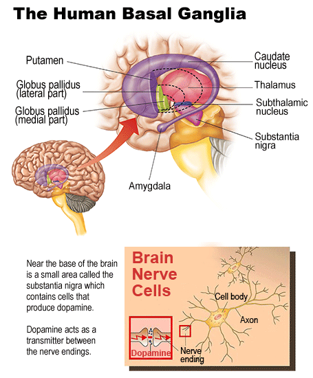
Jay Schneider, one such experimenter, calls it âparkinsonismâ, and writes: âSome monkeys had cognitive deficits and no motor deficits. Other monkeys had full parkinsonism that was produced after short-term high dose MPTP exposure, and some monkeys had full parkinsonism after long-term low-dose MPTP exposure.â[1] ââ¦it is essential to understand that animal models only represent an imperfect replica of human disorders, and this is so for several reasons. First, animal models are generally developed in beings (rodents, non-human primates) that are subjects with behavioural repertoires and anatomical characteristics very different from humans. These species differences are known to play a critical role in the clinical expression as well as in the cellular specificity of the lesions. Second, in addition to these species differences, the time source evolution of the nerve cell generation, which normally evolves over several years in neurodegenerative diseases in humans is for practical reasons being replaced over a much shorter period of time in animal models. â[3] Is this haphazard set of symptoms supposed to be a scientific model? As if to add to the uselessness of the model, the animal model recovers gradually â unlike the chronically afflicted human patients. An expert explains that: âThe best model of PD to date, is theâ¦(MPTP)-lesioned marmosetâ¦.unlike human PD, which is progressive, the neurotoxic damage produced by MPTP is reversible.â [2] The suggestion that gradually recovering monkeys with a condition that causes variable symptoms can be used to study the disease is clearly ludicrous. A further problem which would render animals useless as a method if they werenât already is that communication between animals and human is so limited. Animals canât explain their symptoms, emotions, difficulties in motor functions or what affects them in everyday life. In summary, the animal model is a failure because: Given that animals are a failed method, how has progress been possible against PD? As for the overwhelming majority of illnesses, progress has been due to a combination of study of patients, autopsy, the use of technology and a helping of luck. PD was never really understood until 1960. An Austrian team lead by Oleh Hornykiewicz performed autopsies on human PD patients at the University of Vienna. The knowledge at this point â established by autopsy - was that this very specific, human illness, was caused by a very specific part of the human brain degenerating. The substantia nigra, part of the brain found in the basal ganglia was badly affected. This was never likely to be uncovered in animals, who donât have a comparable basal ganglion. As an expert explains, animal models âdo not reflect the complexities of the human basal ganglion.â[4] The Austrian team discovered that the nigostriatial pathway had degenerated and had very little dopamine. Dopamine regulates movement and emotion, and normally carries most of the nerve signal.
This made perfect sense as PD does affect movement and emotion, and led to tests on brain tissue from PD patients, which confirmed the dopamine deficiency. They went on to discover that nerves containing dopamine die, leaving a deficiency of dopamine. They soon gave patients treatment intended to lead to dopamine production, which caused immediate benefits. As well as revolutionising PD treatment, this enabled better research into other neurological illnesses including epilepsy and schizophrenia. As one expert put it, Hornykiewicz had âfundamentally changed how neuropharmacology is practiced.â[5] Hornykiewicz was never awarded the Nobel Prize for this work, although it was awarded to a team who had followed on from the work pioneered by the Austrians. The award attracted disapproval: 250 neurologists criticised the decision and the decision not to reward Hornykiewicz.[6] This massive discovery is only a step on the way to finding a cure for this terrible disease. The fact that the substantia nigra dies is a symptom, not the cause of the disease, and further knowledge of why it dies needs to be uncovered. For this reason why need more studies into human patients on a detailed level studying the interactions between the substantia nigra and the rest of the body. Autopsies are now possible on a highly detailed level thanks to improvements in microscope technology, and arguably provide the most valuable of any single method in studying nerological illness. Two pathologists wrote âIn recent years, participants in meetings of the American Association of Neuropathologists have heard criticism about the increasing use of animal models to study human neurologic diseaseâ¦. A strong cadre of diagnostic and research neuropathologists believe that only human material can provide relevant answers to many problems about human central nervous system disease. In fact, examination of the data bears out this contention. Of the 185 abstracts presented at the 1985 meeting of the American Association of Neuropathologists, 115 (62%) were presentations of human neuropathology, and an astounding 81 (43%) were based on investigations of human brains at autopsy. Among these autopsy studies were seven presentations of either the first complete description of a newly recognized human disorder, or one of the first complete descriptions of an uncommon human neurologic disease.â[7] Nevertheless, the Austrian discovery made a difference to human patients, as it immediately enabled treatments to enhance dopamine levels. Further human studies showed that Levodopa could be used to stimulate dopamine levels.[8] This is imperfect and becomes less effective over time â the reason for which is understood thanks to study of patients. [9] Another method has been to use the nightshade plant, which decreases levels of acetylcholine, which increases when dopamine is in short supply. Other effects have been discovered by accident â such as the effect of ecstasy or the antiviral Amantadine. Before the Hornykiewicz discovery, surgery was used to control PD by removing both thalami. The thalamus is a two part brain section. This had limited success, but it was in attempting these that a more striking method was discovered.
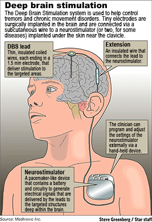
He soon was presented with a patient on whom he couldnât perform a thalamotomy, so tried implanting the electrodes permanently. It was successful, and led to more research, and the eventual approval of the technique around the world.[10] Bernabidâs discovery was a victory for surgical technique, human study, and observation. It owes nothing to animal experiments. This same technique is the one used by Tipu Aziz, an outspoken supporter of the Oxford University animal lab. Aziz has in the past claimed that the treatment of deep brain stimulation given to Parkinson sufferers owes everything to his research on monkey brains and could not have been made in any other way. It is the technique demonstrated by PD patient Mike Robbins. We can be absolutely clear that this technique has nothing to do with animal experiments, and everything to do with the technology, clinical medicine and observation SPEAK and the scientific anti-vivisectionist movement has supported. Deep Brain Stimulation was not discovered by experimenting on non-human animals as the vivisectors would like you to believe. Rather than assisting scientific research in finding cures for human diseases, the use of animals is actually hampering it. We are now in the 21st Century. We have the technological capacity to send space probes to Mars. However, some scientists are still involved in the crude and barbaric practice of vivisection, a practice which dates as far back as the 1600âs. The following section will deal with the new forms of technology that are available to us. These new and innovative types of technology are our best hope in finding cures for human disease. Technology in the modern laboratory fMRI (functional Magnetic Resonance Imaging) This technique identifies the role of different areas of the brain. It does this by detecting higher and lower magnetic susceptibilities in the blood, which indicate whether the blood is newly oxygenated or not. Real time scans are possible which aid treatments such as surgery and are of great value as a diagnostic tool. See http://www.fmrib.ox.ac.uk/fmri_intro/brief.html for more or http://www.dcn.ed.ac.uk/bic/research/structural.asp for a British University applying the technique. MagnetoEncephaloGraphy (or MEG) detects the magnetic fields associated with brain activity without using X-rays. It sends no signals into the brain so is entirely safe. It enables a functional image of the brain to be shown. This helps show what activity the brain is undertaking, and where in the brain this comes from. It helps show where problems (eg epilepsy or migraine) is coming from. See http://www.magres.nottingham.ac.uk/meg/index.phtml for a UK university working at the forefront of this technology. Or see http://www.aston.ac.uk/lhs/research/facilities/meg/faq.jsp and see http://www.aston.ac.uk/lhs/research/groups/nrg/nrg_projects.jsp for some of their valuable work in humans. EIT (Electrical Impedance Tomography), is mobile and cheap. It registers electrical resistance in disease-affected areas. The main benefit is therefore to trace the movement of blood and other fluids. Developments will hopefully lead to this being a cheap, portable method of imaging the brain in full 3-D detail. SPECT (Single Photon-Emission Computed Tomography) enables doctors to build 3D images of the brain by detecting details about the flow of blood. This shows brain function and is vital for detection of illnesses. This is done by radioactive labelling blood. See more at: http://www.amershamhealth-us.com/patient/diaguide/spect.html
PET (Positron emission tomography) scans detect radiation from positrons, and enable a detailed picture of the illness to be constructed. This is vital for patients with brain dysfunction for which the cause has not been determined. See more at http://www.radiologyinfo MRS (Magnetic Resonance Spectroscopy) Enables chemical analysis of the brain without surgery, by distinguishing the chemical nature of the part of the brain being scanned. This is done by detecting the magnetic resonance in that part of the brain and analysing the data this shows. http://www.ness-foundation EROS Uses lasers which can pass through the skull, to image the brain. They are fired from dozens of different directions at once, and the technique measures differences in the way they reflect. The differences are caused by the fluid in the brain cells, and reveal vital information about the condition of the different parts of the brain. http://www.sciencentral.com/articles/view.php3? TMS (Transcranial magnetic stimulation) stimulates or calms parts of the brain using magnetic impulses. Higher frequencies stimulate, lower ones calm. This enables doctors to calm brain areas and assess the affect on symptoms, therefore identifying brain areas linked with specific illnesses. Long-term imbalances in the brain can be identified. http://www.ucl.ac.uk/news/news-articles/06080702 Without autopsies, the progress in neurology would be almost non existent. This method has focused on real patients and the real nature of their brains, and full records of their condition have been compared with the findings. As microscopes become more powerful, the method becomes more effective. In vitro study involves studying human brain tissue and understanding the chemical interactions and detailed information about the biology of it. It also compares healthy tissue with unhealthy tissue to show the difference, and has already helped enormously with development of treatments. Computer modelling is becoming more advanced each year. With the information from autopsy, in vitro studies and technological imaging, knowledge of the brain and activity in it is more detailed than ever. The healthy brain, and the brain afflicted by illnesses can now be modelled and the complex interactions can be modelled. The human brain is unlike any other, with additional areas, different proportions, and is organised differently. The only way the function of the human brain has been understood has been through clinical (human) study. Studies have shown that the same areas in different animal and human brains play different roles as well: damage to the corresponding parts of monkey and human brains has been shown do cause different symptoms.[11] In the early 1800s the speech centres of the brain were located through autopsies and observing patients â work which would have been impossible through vivisection as animals lack the same speech process more obviously than they lack other processes.[12] Research into human brain function is only really possible through studying humans â either in life or at post mortem. As a recognised neurologist explains: âThe study of the brain, if it is to bear fruit, must be made on man, i.e. at the bedside and in the post-mortem theatre; â¦The utmost that can be learned from experiments on the brains of animals is the topography of the animalâs brain, and it must still remain for the science of human anatomy and clinical investigation to enlighten us in regardâ¦of our own species; and in fact, it is from the department of clinical investigation and post-mortem study that so far all of our best brain localizations have been secured.â[13] Other illnesses affecting the brain Human illness cannot be researched by using animals. The evidence that proves this played a major part of the decision not to build a similar lab in Girton, attached to Cambridge University. The evidence was written by a doctor and can be viewed by going to: http://www.vivisection-absurd.org.uk/xprimate.html and selecting âpart 2â. Some of the other illnesses of the brain are also claimed to be advanced through animal study, but as with PD, progress has only been possible through human study. Multiple Sclerosis MS is caused by the immune system attacking itself, and the cause is not yet known. Animal models have misled experimenters about how MS progresses[14] and have symptoms and patterns of damage to the brain that are not at all in line with human experience.[15] They have also failed to provide treatments of value. âTime after time, researchers have discovered new ways to cure laboratory rats of experimental induced encephalomyelitis, the murine model of MS, only to face obstacles in bringing the treatment to humans.â[16] Treatments developed on animals include Tumor necrosis factor (which has the opposite effect in humans) [17], Copaxone (which came with numerous side effects)[18], and injected immunoglobins, which were no more effective than placebos [19]. Peptide ligand formulas trials were abandoned as patients nearly died.[20] Most research into MS has been based on clinical research and culture work using T-cell lines and cells taken from individual patients.[21] Alzheimer's Disease Dr Alois Alzheimer first identified AD using microscopes and autopsy. The lack of mental function is caused by protein becoming uncontrolled and forming protein deposits in the brain now known as neurofibrillary tangles.[22] The particular protein involved (tau) was identified in mice, but altering the mouseâs tau did not even cause any symptoms similar to AD, despite enormous efforts and resources.[23] The first major breakthrough was the discovery that AD patients lacked acetylcholine, which helps neurons communicate. This was discovered through autopsy.[24] Animal models have proved frustrating and unproductive: âThe full spectrums of the biochemical and pathological abnormailities characterized by AD have not been found to occur spontaneously in any animal species other than humanâ¦â[25] Another explains: âThere is no good animal model for the disease process characterized by a loss of cognitive functions and memory decline.â[26] While human studies have enabled advances, especially in the knowledge of the genetic implications of AD, a medical journal editorial criticising animal models points out that the first discovered characteristic of AD is not present in animal models: âMore problematically, these animals do not develop neurofibrillary tangles or show significant neurodegeration.â[27] Epilepsy In a speech at the International Symposium of April 25 1987, Zurich, Dr Med. Bernhard Rambeck, Director of the Biochemistry Department of the Society for Epilepsy Research in Bielefield-Bethel, West Germany stated: The first accurate description of epilepsy was by John Hughlings Jackson. Long before electroencephalograms (EEGs) he correctly described the illness as one caused by abnormal electrical discharges in the brain. This was based on observing patients, although now, MRIs can show which part of the brain is causing the seizures and how the illness is progressing. Progress has enabled types of epilepsy to be identified: âThe detailed models for focal interictal discharges arose largely from experiments on brain slices in vitro [studied after death], combined with computer simulations.â [28] An evaluation of modern epilepsy research by vivisection supporters recently identified the most important methods as: â¢Brain imaging methods, especially MRIâ¢Surgical technique and the ability to detect opportunities for surgery â¢Molecular genetics Animal experiments were not mentioned.[30] Attempts to cause an animal model by inducing birth defects in animals were based on the discovery that substance abuse in humans is linked to epilepsy. It didnât work.[31] The discovery of treatments has been mainly due to discovering the effects medicines have had on epilepsy when intended for another condition. âOverwhelmingly, discovery of the old and a number of the new AEDs [antiepileptic drugs] came from serendipity [chance].â[32] Unsurprisingly, animal experiments are seen as a poor method of studying human illness and are rapidly becoming less respected. As two pathologists explain: The idea that animal models would enable this is dismissed: Although dogs can naturally develop epilepsy, comparing human and dog patients is not viable due to the way the drugs are handled by the bodies. Vigabatrin treats epileptic dogs, but when given to human was related to severe vision damage and has led to large court cases.[34] The claim by animal experimenters that their practice has been invaluable is false. It is also a claim which other researchers feel is detrimental to the advancement of science. Concern about the preoccupation with animal research is becoming more prevalent. The evidence that animal experiments are bad science and hamper the progress of human medicine is overwhelming and more doctors are coming to the conclusion that vivisection is a deeply flawed scientific practice. Vivisectors want to continue with animal experiments not because itâs the best form of research but because it keeps them in their chosen career. Itâs vitally important that for those people suffering from terrible illnesses, that the fraud is stopped now. See the following websites for more information: References |
Home | About SPEAK | Make A Donation | Resources | Links | News Archive | Contact Us | Search | Demo Diary  Disclaimer: The information on this website is for the purpose of legal protest and information only. It should not be used to commit any criminal acts or harassment. SPEAK Campaigns � speakcampaigns.org. 2004 |